Compact Bone Diagram Lacunae : Compact Bone Structure Biology Dictionary - Between the rings of matrix, the bone cells (osteocytes) are located in spaces called lacunae.
Compact Bone Diagram Lacunae : Compact Bone Structure Biology Dictionary - Between the rings of matrix, the bone cells (osteocytes) are located in spaces called lacunae.. Thus, the lamellar pattern as well as the lacunae size differ between trabecular and cortical bone. Explain the role of bone salts and the organic matrix in making bone both hard and flexible. The remainder is cancellous bone, which has a spongelike appearance with numerous large spaces and is found in the. The lamellae are difficult to distinguish, but the haversian canals and the lacunae can be easily. The lacunae are thought to be caused by erosion of the bone by the osteoclasts enzymes.
Usually found in long bones of the body, it consists of units called. The compact bones form the hard exterior of the bones, whereas the spongy bones have several pores that are filled with nerves and blood vessels. The marrow in these images is red marrow. Each haversian canal generally contains one or two capillaries and many nerve fibres.the channels are formed by concentric layers called lamellae, which are approximately 50 µm in diameter.the haversian canals surround blood vessels and nerve cells throughout bones and communicate with osteocytes (contained in spaces within the dense bone matrix called lacunae) through connections. The organic components are present, and the section is stained with hematoxylin and eosin.
Structure Of Bone Tissue Bone Structure Anatomy Components Of Bones Youtube from i.ytimg.com Identify structures and functions of the microscopic structure of compact and spongy bone 3. The marrow in these images is red marrow. The remainder is cancellous bone, which has a spongelike appearance with numerous large spaces and is found in the. A diagram of the femur showing a cut through the compact bone into the medullary cavity. Compact bone, as opposed to spongy bone, is made of cylindrical units, called osteons, that are tightly formed together. The organic components are present, and the section is stained with hematoxylin and eosin. Compact bone makes up 80. An enlarged view of an osteon showing the ostecytes within lacunae and the concentric lamellae.
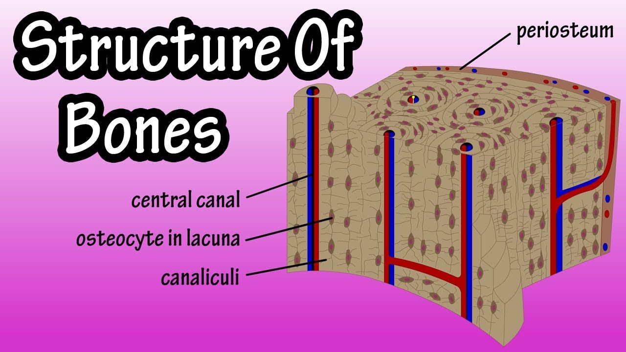
The osteon consists of a central canal called the osteonic (haversian) canal, which is surrounded by concentric rings (lamellae) of matrix.
I.e., slide 56, however, is not a ground section. Compact bone, also called cortical bone, dense bone in which the bony matrix is solidly filled with organic ground substance and inorganic salts, leaving only tiny spaces (lacunae) that contain the osteocytes, or bone cells.compact bone makes up 80 percent of the human skeleton; Human bone generally comprises osseous tissue, an outer coating called a periosteum, and bone marrow.the two main structural components typically include spongy bone on the interior, with an outer layer of compact bone. (on textbook page diagrams note only highlighted labels) compact bone. Learn vocabulary, terms, and more with flashcards, games, and other study tools. Compact bone makes up 80. Compact bone forms the surface of all bones. Learn vocabulary, terms, and more with flashcards, games, and other study tools. Usually found in long bones of the body, it consists of units called. Iv.3 third level trabecular bone structure Compact bone, as opposed to spongy bone, is made of cylindrical units, called osteons, that are tightly formed together. Compact bone structure diagram quizlet from o.quizlet.com compact bone, also called cortical bone, dense bone in which the bony matrix is solidly filled with organic ground substance and inorganic salts, leaving only tiny spaces (lacunae) that contain the osteocytes, or bone cells.compact bone makes up 80 percent of the human skeleton; The osteon consists of a central canal called the osteonic (haversian) canal, which is surrounded by concentric rings (lamellae) of matrix.
Learn vocabulary, terms, and more with flashcards, games, and other study tools. (on textbook page diagrams note only highlighted labels) compact bone. The organic components are present, and the section is stained with hematoxylin and eosin. A diagram of the femur showing a cut through the compact bone into the medullary cavity. The tissue level of organization.
Bme 332 Bone Structure Function from umich.edu Canaliculus compact bone haversian canal lacuna lamellae osteocyte osteon ii spongy bone check answer this problem has been solved! Like compact bone, spongy bone, also known as cancellous bone, contains osteocytes housed in lacunae, but they are not arranged in concentric circles. The marrow in these images is red marrow. The organic components are present, and the section is stained with hematoxylin and eosin. Each haversian canal generally contains one or two capillaries and many nerve fibres.the channels are formed by concentric layers called lamellae, which are approximately 50 µm in diameter.the haversian canals surround blood vessels and nerve cells throughout bones and communicate with osteocytes (contained in spaces within the dense bone matrix called lacunae) through connections. Start studying anatomy compact bone diagram. An osteocyte within a lacuna. Compact bone, as opposed to spongy bone, is made of cylindrical units, called osteons, that are tightly formed together.
The lamellae are difficult to distinguish, but the haversian canals and the lacunae can be easily.
The compact bones form the hard exterior of the bones, whereas the spongy bones have several pores that are filled with nerves and blood vessels. Bone marrow is present (it is a kind of haematopoietic tissue from which all blood cells are made). Compact bone forms the surface of all bones. The lamellae are difficult to distinguish, but the haversian canals and the lacunae can be easily. In long bones, as you move from the outer cortical compact bone to the inner medullary cavity, the bone transitions to spongy bone. Compact bone makes up 80. 0 0000 a shoutout is a way of letting people know of a. Anatomy of a long bone proximal epiphysis diaphysis distal epiphysis compact bone spongy bone medullary cavity. Noun plural lacunae luh kyoo nee ləˈkyu ni lacunas. The trabeculae are only a few cell layers thick. Between the rings of matrix, the bone cells (osteocytes) are located in spaces called lacunae. An enlarged view of an osteon showing the ostecytes within lacunae and the concentric lamellae. These in turn are derived from the bone marrow.
Between the rings of matrix, the bone cells (osteocytes) are located in spaces called lacunae. Compact bone makes up 80. Learn vocabulary, terms, and more with flashcards, games, and other study tools. A structural unit of compact bone consisting central haversian canal. A diagram of the femur showing a cut through the compact bone into the medullary cavity.
Bone Tissue Amboss from media-us.amboss.com I.e., slide 56, however, is not a ground section. The arrangement of the osteons within the diaphysis of the bone. The trabeculae are only a few cell layers thick. The compact bones form the hard exterior of the bones, whereas the spongy bones have several pores that are filled with nerves and blood vessels. Compact bone structure diagram quizlet from o.quizlet.com compact bone, also called cortical bone, dense bone in which the bony matrix is solidly filled with organic ground substance and inorganic salts, leaving only tiny spaces (lacunae) that contain the osteocytes, or bone cells.compact bone makes up 80 percent of the human skeleton; Section is composed of compact bone exactly like the compact bone on slide 54. About press copyright contact us creators advertise developers terms privacy policy & safety how youtube works test new features press copyright contact us creators. Explain the role of bone salts and the organic matrix in making bone both hard and flexible.
The organic components are present, and the section is stained with hematoxylin and eosin.
Between the rings of matrix, the bone cells (osteocytes) are located in spaces called lacunae. I.e., slide 56, however, is not a ground section. The spaces between the trabeculae contain red or yellow marrow, depending on a person's age and on which bone it is. Each haversian canal generally contains one or two capillaries and many nerve fibres.the channels are formed by concentric layers called lamellae, which are approximately 50 µm in diameter.the haversian canals surround blood vessels and nerve cells throughout bones and communicate with osteocytes (contained in spaces within the dense bone matrix called lacunae) through connections. (on textbook page diagrams note only highlighted labels) compact bone. Identify structures and functions of the microscopic structure of compact and spongy bone 3. In osteoclast on the bones surface called howship lacunae. The compact bones form the hard exterior of the bones, whereas the spongy bones have several pores that are filled with nerves and blood vessels. An osteocyte within a lacuna. The tissue level of organization. In long bones, as you move from the outer cortical compact bone to the inner medullary cavity, the bone transitions to spongy bone. A structural unit of compact bone consisting central haversian canal. Compact bone makes up 80.
To know the structures of a synovial joint and a symphysis joint (intervertebral disc) compact bone diagram. These are active participants of blood supply.


0 Komentar